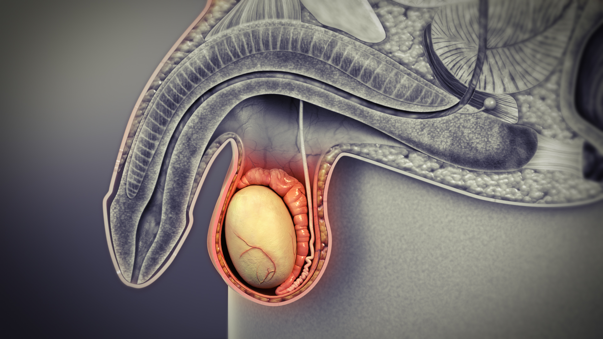
The testes are two oval-shaped organs which are part of the male reproductive system, and are also known as ‘testicles’. They are enclosed in the scrotum, which is a sac like structure of skin that hangs outside the body in the pelvic region close to upper thighs.
Anatomy and functions
The most important function of the testes is to produce and store sperms. They are also vital for forming testosterone and other male hormones namely androgens.
The ovular shape of testes is due to the tissues called as lobules. It consists of coiled tubes surrounded by dense connective tissues.
Seminiferous tubules
These are the coiled tubes that form the bulk of each testis. The cells and tissues in the tubules are responsible for the process of forming sperms.
These tubules are lined with the epithelium. This layer consists of Sertoli cells that help in the development of hormones that generate sperm.
Leydig cells are the tissues next to the tubules, which produce male hormones namely testosterone and other androgens.
Rete testis
Once the sperm is formed in the seminiferous tubules, the sperm cells travel towards the epididymis via the rete testis. The rete testis aids in mixing sperm cells around in the fluid secreted by the Sertoli cells.
The sperms can’t move before they can get to the epididymis. Millions of small projections known as microvilli, help move sperm along to the efferent tubules.
Efferent ducts
The efferent ducts are the tubes that connect the rete testis to the epididymis. The sperm cells until mature and ready for ejaculation, are stored in the epididymis.
Hair-like projections called cilia, form the lining of these ducts. Along with a layer of smooth muscle, cilia help move the sperm into the epididymis.
The efferent ducts absorb most of the fluid that helps to move the sperm cells also. This accounts for a greater sperm concentration in the ejaculate fluid.
Tunica: Vasculosa, albuginea, and vaginalis
The testicles are surrounded by several layers of tissues. These are as below:
- Tunica Vasculosa
- Tunica Albuginea
- Tunica Vaginalis
Tunica Vasculosa is the first thin layer of blood vessels which protects the tubular interior of each testicle from further layers of tissue.
Tunica Albuginea is the next layer which is a thick, protective layer made of densely packed fibers that further shield the testes.
Tunica Vaginalis is the outermost layers of tissue. It consists of three layers as below:
- Visceral layer
- Cavum vaginale
- Parietal layer
Conditions of the testes
- Varicocele: In this condition there is swollen vein(s) from the testes which most commonly affects the left side. The testicles are usually normal.
- Hydrocele testis: In this condition, there is swelling around the testes due to accumulation of clear liquid within a membranous sac. The testicles are usually normal.
- Testicular cancer and other neoplasms
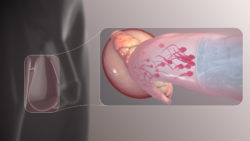
- Spermatocele: In this disease, there is a retention cyst of a tubule of the rete testis or epididymis’ head, swollen with barely watery fluid that has spermatozoa
- Endocrine disorders: These disorders may also affect testis’ size and function.
- Bell-clapper deformity is a deformity in which there is not attachment of testes to the scrotal walls. It can rotate without restriction on the spermatic cord within the tunica vaginalis. It is the most usual cause of testicular torsion.
- Orchitis inflammation of the testes
- Epididymitis: In this condition there is a painful inflammation of the epididymis. It is often caused by the bacterial infection however sometimes of unknown origin.
- Anorchia: In this condition there is absence of one or both testicles.
- Cryptorchidism: This happens in cases where the testes does not descend into the scrotum of the infant boy and a few more conditions.
Disclaimer: The information in no way constitutes, or should be construed as medical advice. Nor is the above article an endorsement of any research findings discussed in the article an endorsement for any of the source publications.
Sources-
- https://www.healthline.com/human-body-maps/testis
- https://en.wikipedia.org/wiki/Testicle
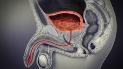
The bladder is an expandable saclike organ that contracts once its empty, just like the stomach. Its inner lining constricts into the folds and expands out to accommodate liquid.
Read More..
Finally it looks like a male contraceptive method that is non-surgical, non-occlusive and reversible is on its way.
Read More..